Arthroscopy
Arthroscopy in Los Angeles
A small camera can examine joints such as the elbow or shoulder and guide selected treatment. Examination and imaging determine whether arthroscopy or another approach is appropriate.

Procedure overview
Arthroscopy uses a small camera and dedicated instruments to examine the inside of a joint. It may be used to diagnose or treat selected joint conditions in dogs and cats after examination and imaging review.
Arthroscopic applications
- Elbow: Evaluation and treatment of selected elbow dysplasia conditions and osteochondral lesions.
- Shoulder: Evaluation of osteochondral lesions, instability or biceps-tendon concerns.
- Stifle: Inspection of selected meniscal and intra-articular concerns, including cases planned with another orthopedic procedure.
- Other joints: Carpal, tarsal and other small-joint evaluation in selected patients where anatomy and instrument access allow.
Is arthroscopy appropriate?
Arthroscopy may be considered when examination and diagnostic imaging suggest an intra-articular problem that would benefit from direct visualization or treatment. Patient size, joint anatomy, condition and the available alternatives are reviewed before an approach is recommended.
What the procedure may involve
Under general anesthesia, the joint is accessed with a small camera and dedicated instruments. Depending on the joint and diagnosis, the plan may involve direct inspection, treatment of selected osteochondral or dysplasia-related lesions, assessment of a tendon or meniscus, or removal of selected intra-articular material. Arthroscopy may also be planned alongside another orthopedic procedure when clinically appropriate.
Recovery planning
Pain management, activity restriction, rehabilitation and recheck timing depend on the joint and procedure performed. The clinic provides a plan specific to the patient and coordinates relevant records with a referring veterinarian when applicable.
- Approach
- Camera-assisted joint access
- Clinical use
- Selected joint conditions
- Anesthesia
- General anesthesia
- Planning
- Imaging and exam review
Procedure recommendations depend on physical exam findings, diagnostics, anatomy, condition severity and anesthetic safety. Dr. Antonio Pedraza reviews each case individually.
Clinical background and sources
These resources explain the general techniques and care considerations. Your veterinary team determines how they apply to your companion and the proposed procedure.
American College of Veterinary Surgeons
Canine Elbow DysplasiaElbow dysplasia, assessment and treatment options, including selected arthroscopic procedures.
American Animal Hospital Association
2020 AAHA Anesthesia and Monitoring Guidelines for Dogs and CatsIndividual pre-anesthetic assessment, monitoring and the transition into recovery.
Clinical imagery
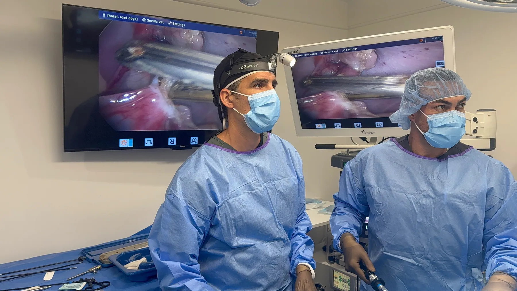
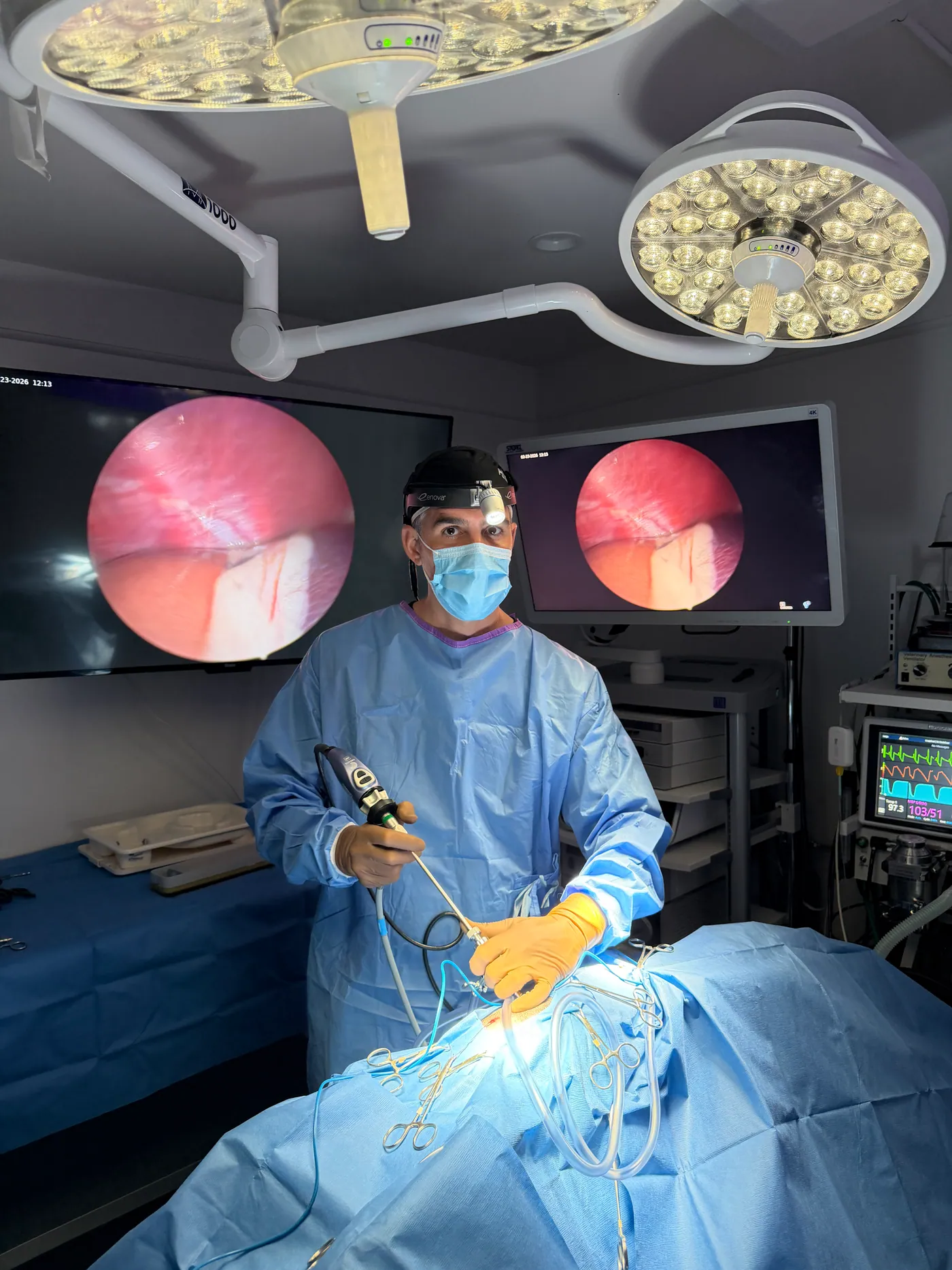
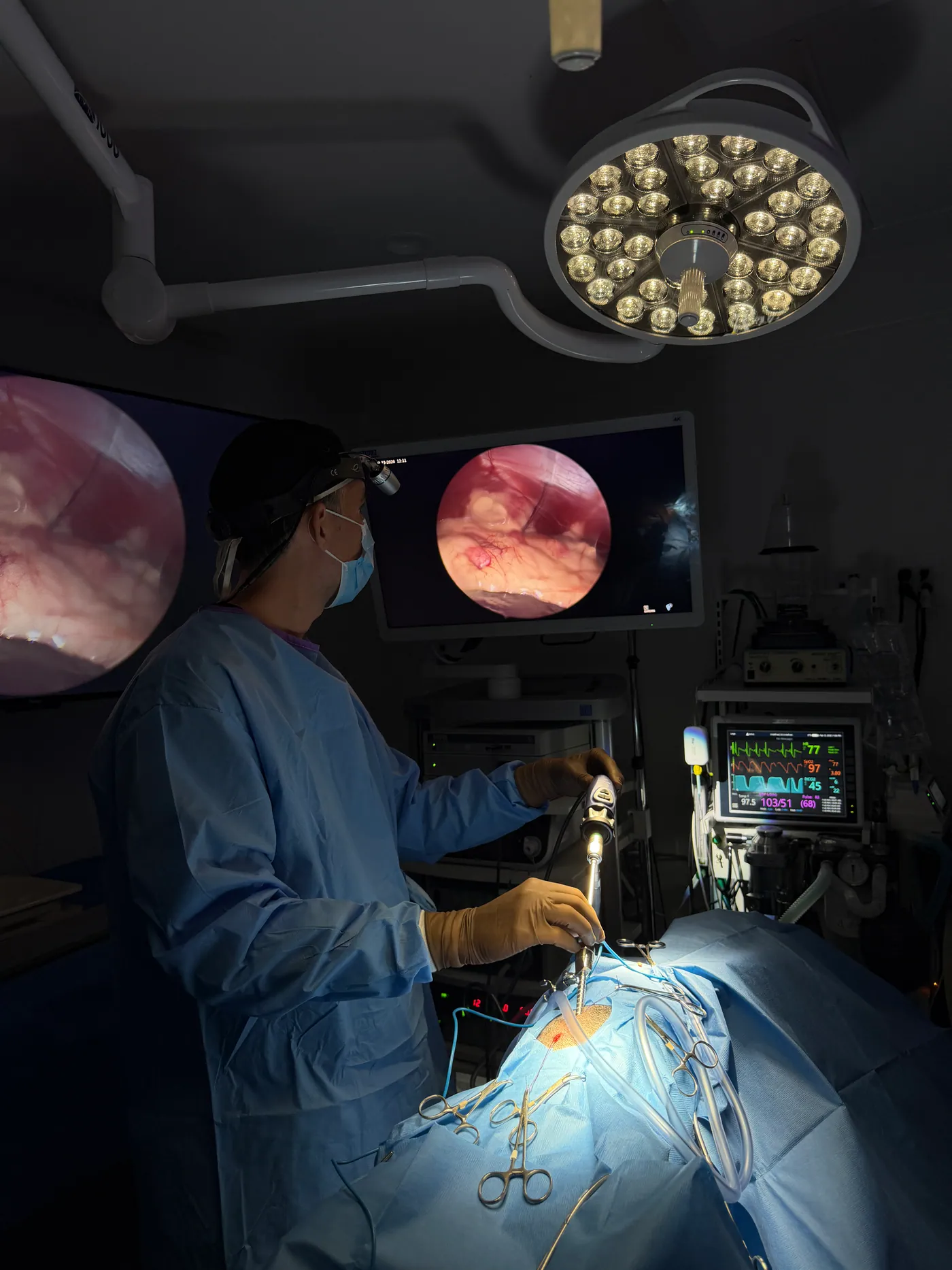
Surgical environment and visualization equipment
Photographs of the surgical environment and visualization equipment at the clinic.
3 clinical views
Santa Monica · Greater Los Angeles
Advanced care from a local clinical team
Los Angeles Laparoscopy Vet at Sevilla Veterinary Hospital is located at 2116 Main Street in Santa Monica. The clinic serves companion animals from Santa Monica, Los Angeles, West Los Angeles, Venice, Brentwood, Culver City, Beverly Hills, Pacific Palisades, Marina del Rey.
Los Angeles Laparoscopy Vet at Sevilla Veterinary Hospital
2116 Main Street
Santa Monica, CA 90405
Next step
Ready to discuss your companion's care?
Speak with our team about the available approaches and whether a minimally invasive procedure may be appropriate after individual evaluation.