VITOM Microscope
VITOM Surgical Microscope
An exoscope magnifies a surgical site from outside the body. Unlike a scope inserted into a body cavity, VITOM supports external viewing while the surgeon works.

Procedure overview
The VITOM surgical visualization system is an exoscope positioned above the surgical field. It displays a magnified, illuminated view on a high-definition monitor while leaving working space around the patient.
How the VITOM system works
A telescopic camera is mounted on a stable arm outside the body and directed toward the operative field. The surgical team can view the same magnified image on a monitor, which can support shared orientation and documentation during selected procedures.
Where magnified visualization may help
- Detailed viewing: Magnification can make small structures easier for the surgical team to see.
- Shared monitor view: Team members can follow the same operative image.
- External positioning: The camera remains outside the patient and does not occupy the surgeon's direct viewing position.
- Selected applications: The system may be used for spinal, eyelid, ear, oncologic or other procedures where an exoscopic view adds clinical value.
The role and limits of the system
VITOM is a visualization tool rather than a procedure by itself. The underlying surgery, anesthesia, recovery and expected course depend on the condition being treated.
The exoscope may support detailed viewing, team orientation and documentation, but it does not determine whether an operation is minimally invasive and does not replace the clinical judgment required to choose an access route or procedure.
- Approach
- External exoscopic visualization
- Clinical use
- Magnified surgical viewing
- Anesthesia
- Depends on the procedure
- Planning
- Used when visualization adds value
Procedure recommendations depend on physical exam findings, diagnostics, anatomy, condition severity and anesthetic safety. Dr. Antonio Pedraza reviews each case individually.
Clinical background and sources
These resources explain the general techniques and care considerations. Your veterinary team determines how they apply to your companion and the proposed procedure.
KARL STORZ — device manufacturer
Veterinary Imaging Solutions: VITOMManufacturer description of external magnified visualization for open veterinary surgery; not evidence of a patient outcome.
American Animal Hospital Association
2020 AAHA Anesthesia and Monitoring Guidelines for Dogs and CatsIndividual pre-anesthetic assessment, monitoring and the transition into recovery.
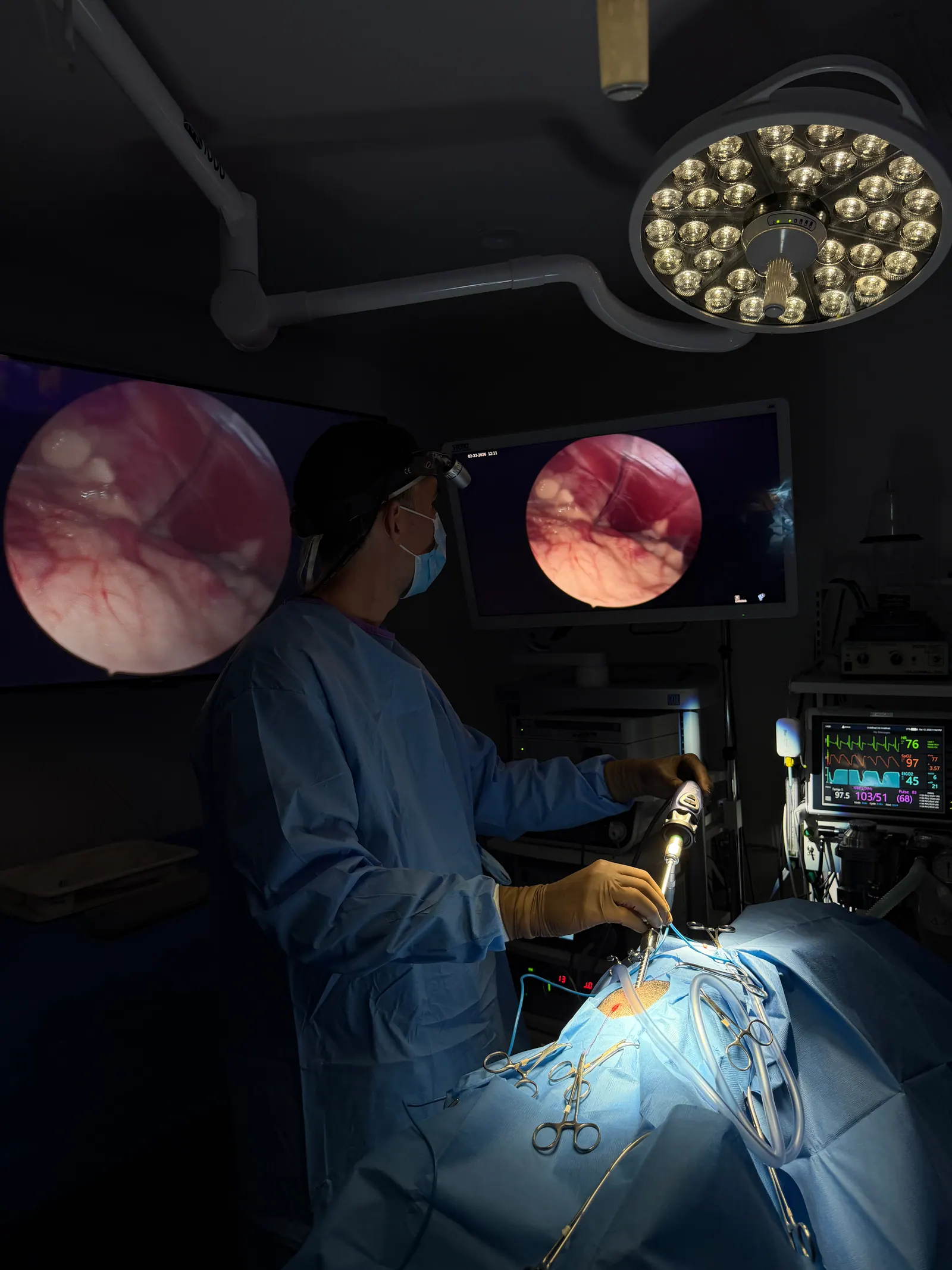
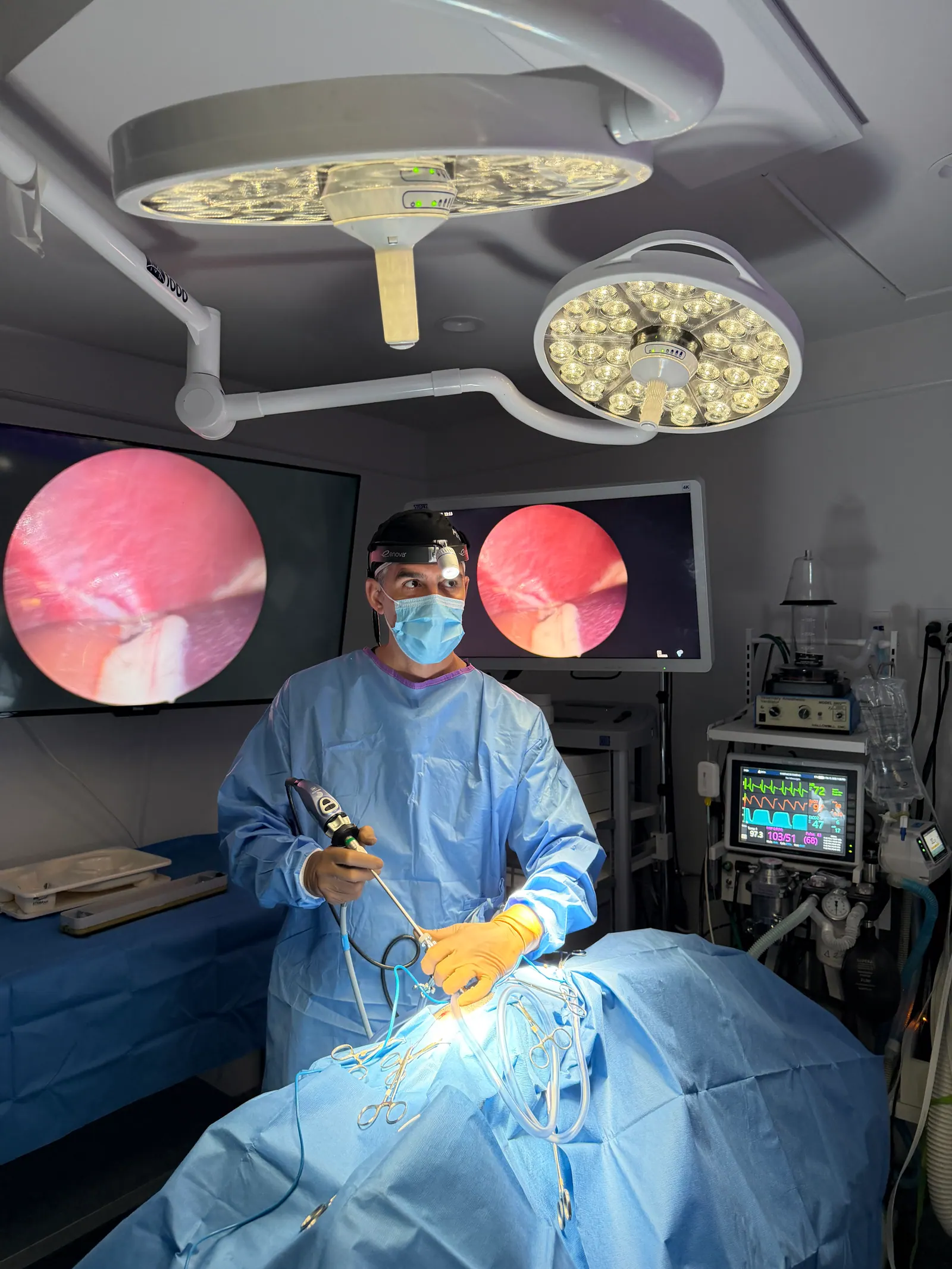
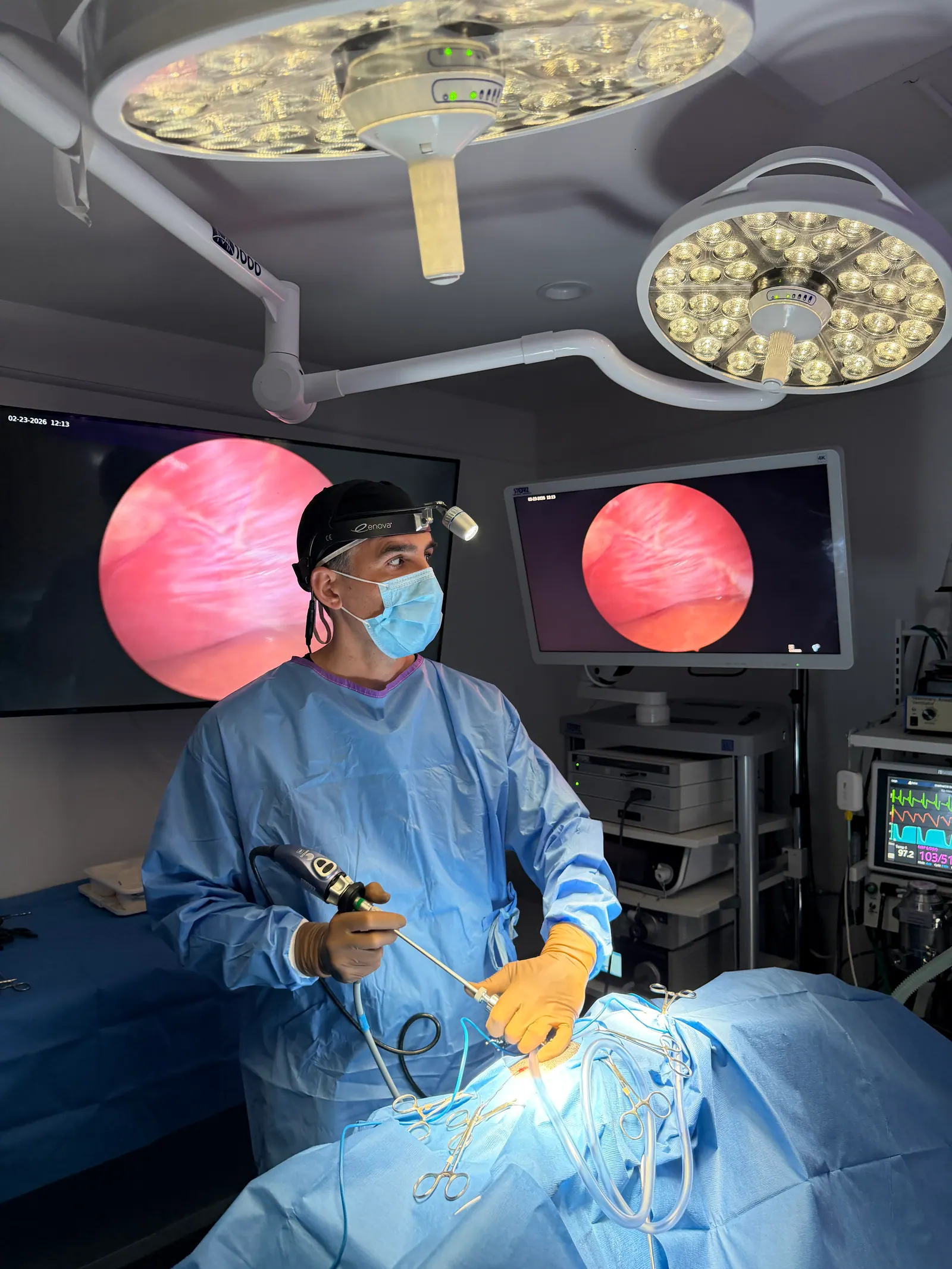
Clinical imagery
Surgical environment and visualization equipment
Photographs of the surgical environment and visualization equipment at the clinic.
3 clinical views
Santa Monica · Greater Los Angeles
Advanced care from a local clinical team
Los Angeles Laparoscopy Vet at Sevilla Veterinary Hospital is located at 2116 Main Street in Santa Monica. The clinic serves companion animals from Santa Monica, Los Angeles, West Los Angeles, Venice, Brentwood, Culver City, Beverly Hills, Pacific Palisades, Marina del Rey.
Los Angeles Laparoscopy Vet at Sevilla Veterinary Hospital
2116 Main Street
Santa Monica, CA 90405
Next step
Ready to discuss your companion's care?
Speak with our team about the available approaches and whether a minimally invasive procedure may be appropriate after individual evaluation.