Laser & Energy
Laser & Energy Procedures
Selected airway and soft-tissue procedures may use laser or energy devices. The tissue, anatomy and clinical goal determine which device and approach are appropriate.

Procedure overview
Laser and advanced energy devices deliver controlled energy to cut, ablate, coagulate or seal tissue. The device and settings are selected according to the tissue, access route and clinical objective.
Laser surgery
A surgical laser uses focused light energy as an alternative or complement to other instruments. It may be considered for selected skin, oral, upper-airway and endoscopic procedures after evaluation.
- Skin and mass procedures: Selected cyst, wart and mass removals.
- Soft-palate and oral procedures: Selected upper-airway and oral tissue procedures.
- Endoscopic laser applications: Selected nasal, urinary, esophageal or airway procedures where laser access is appropriate.
Endoscopic laser applications
A laser may be delivered through or alongside an endoscope when the access route, tissue and clinical goal make that appropriate. Applications include selected nasal tissue procedures, scar-tissue treatment associated with an esophageal narrowing, urinary stone fragmentation and selected urinary or airway applications. Availability and candidacy require individual review; the same device is not appropriate for every lesion or location.
Advanced energy devices
- Electrocautery and radiosurgery: Energy-based cutting and coagulation during selected procedures.
- Vessel-sealing devices: Instruments designed to seal vessels during open or laparoscopic surgery.
- Ultrasonic instruments: Mechanical and ultrasonic energy for selected dissection and sealing tasks.
- Coblation: Radiofrequency energy in saline for selected soft-tissue applications.
Potential advantages and limitations depend on the procedure, device, tissue and individual patient. Dr. Pedraza discusses why a particular instrument is being considered as part of the surgical plan.
Planning and limitations
Laser and energy devices describe how tissue is handled, not the complete procedure. The underlying diagnosis determines whether imaging, biopsy, open access, endoscopic access or another treatment is needed. Anesthesia, discharge and recovery instructions therefore vary with the operation rather than with the device name alone.
- Approach
- Focused energy device
- Clinical use
- Selected tissue procedures
- Anesthesia
- Depends on the procedure
- Planning
- Device chosen by clinical goal
Procedure recommendations depend on physical exam findings, diagnostics, anatomy, condition severity and anesthetic safety. Dr. Antonio Pedraza reviews each case individually.
Clinical background and sources
These resources explain the general techniques and care considerations. Your veterinary team determines how they apply to your companion and the proposed procedure.
American College of Veterinary Surgeons
Brachycephalic SyndromeAirway anatomy, clinical signs, evaluation and surgical options for brachycephalic animals.
American Animal Hospital Association
2020 AAHA Anesthesia and Monitoring Guidelines for Dogs and CatsIndividual pre-anesthetic assessment, monitoring and the transition into recovery.
Specific laser & energy procedures
Brachycephalic Airway Surgery (BOAS) for Dogs in Los Angeles
Does your French Bulldog, Pug or Bulldog snore, snort or tire fast? Learn about brachycephalic airway syndrome (BOAS) and minimally invasive laser airway surgery in Santa Monica with Dr. Pedraza.
Soft Palate & Nostril Surgery for Flat-Faced Dogs
Soft palate and nostril surgery are the most common corrections for brachycephalic (BOAS) breathing problems. Learn how laser-assisted airway surgery works, with Dr. Pedraza in Santa Monica.
Clinical imagery
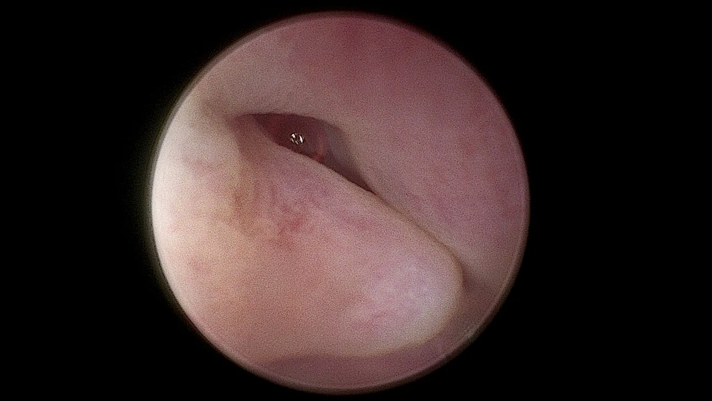
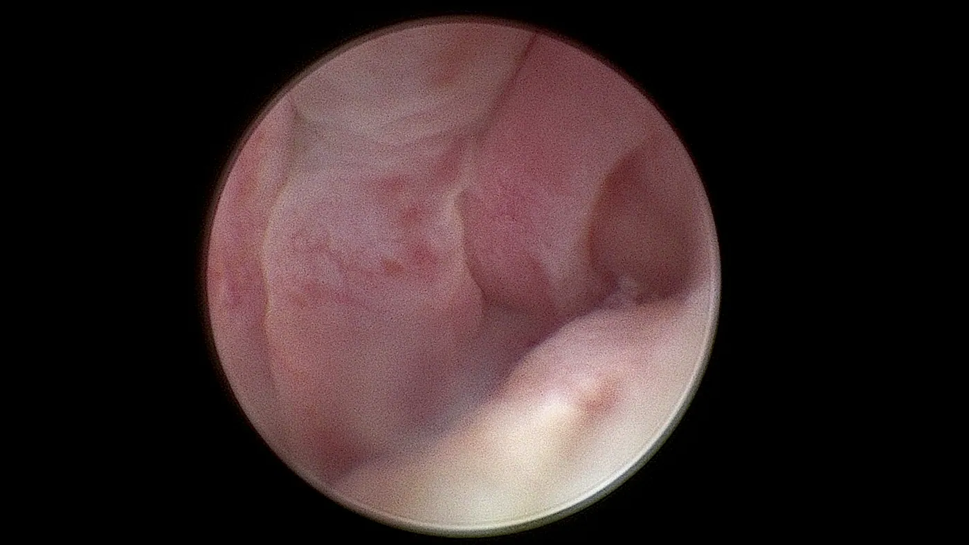
Clinical anatomy and procedure imagery
Surgical and endoscopic views of anatomy and procedure context. Findings vary by patient.
2 clinical views + surgical footage
View the clinical imagery2 views · internal anatomy
View clinical footage Silent video · internal anatomy
Rhinoscopy assessment showing inflammatory changes and polyps.
Visual description: Silent internal camera footage. The camera moves through pink nasal tissue, then approaches rounded, uneven surfaces with yellow material and fluid. The angle and reflections change as the camera moves closer. This is a short excerpt of the assessment.
Santa Monica · Greater Los Angeles
Advanced care from a local clinical team
Los Angeles Laparoscopy Vet at Sevilla Veterinary Hospital is located at 2116 Main Street in Santa Monica. The clinic serves companion animals from Santa Monica, Los Angeles, West Los Angeles, Venice, Brentwood, Culver City, Beverly Hills, Pacific Palisades, Marina del Rey.
Los Angeles Laparoscopy Vet at Sevilla Veterinary Hospital
2116 Main Street
Santa Monica, CA 90405
Next step
Ready to discuss your companion's care?
Speak with our team about the available approaches and whether a minimally invasive procedure may be appropriate after individual evaluation.